


Description
GLUT-1
All names and symbols: solute carrier family 2 member 1; solute carrier family 2, facilitated glucose transporter member 1; Glucose transporter type 1, erythrocyte/brain; HepG2 glucose transporter; GLUT-1; GLUT1; CSE; DYT17; DYT18; DYT9; EIG12; GLUT; GLUT1DS; HTLVR; PED; SDCHCN; SLC2A1
| Host species | Rabbit |
| Clonality | Recombinant |
| Isotype | IgG |
| Confirmed species reactivity | Human |
| Confirmed applications | IHC |
| Aeonian Rating® | 92 |
| RRID | AB_2920807 |
Immunohistochemistry (IHC):
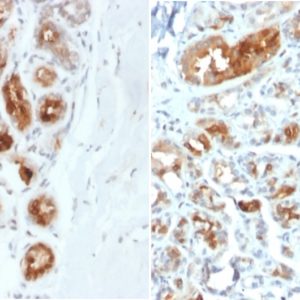
AE00369 to GLUT-1 was successfully used to stain plasma membranes of endothelial cells in human cerebrum, erythrocytes in human liver and trophoblasts in human placenta sections. Recommended concentration: 1-3ug/ml

Formaldehyde-fixed, paraffin-embedded human cerebrum stained with GLUT-1 Rabbit Recombinant Antibody AE00369 at 1-2ug/ml for 60 minutes at 37°C. Epitope retrieval: Autoclave at 121°C, pH7.8 for 5 min followed by 20 min cooling. DAB staining by HRP polymer.

Formaldehyde-fixed, paraffin-embedded human liver stained with GLUT-1 Rabbit Recombinant Antibody AE00369 at 1-2ug/ml for 60 minutes at 37°C. Epitope retrieval: Autoclave at 121°C, pH7.8 for 5 min followed by 20 min cooling. DAB staining by HRP polymer.

Formaldehyde-fixed, paraffin-embedded human placenta stained with GLUT-1 Rabbit Recombinant Antibody AE00369 at 1-2ug/ml for 60 minutes at 37°C. Epitope retrieval: Autoclave at 121°C, pH7.8 for 5 min followed by 20 min cooling. DAB staining by HRP polymer.
Tissue Microarray Validation:
Normal tissues
Negative tissues containing significant positive erythrocytes are categorised as positive
| Strong positive | Weak to moderate | No staining at all |
|---|---|---|
| adrenal gland (endothelium) | appendix mucosa (follicular dendritic cells) | aorta endothelium |
| bone marrow (erythroblasts) | ectocervix (epithelium) | appendix muscular wall |
| cerebellum (endothelium) | endometrium (epithelial and stromal cells) | colon muscular wall |
| cerebrum (endothelium) | epididymis (erythrocytes) | endocervix |
| colon mucosa (endothelium, erythrocytes) | esophagus squamous epithelium | parotid gland |
| duodenum Brunner gland (erythrocytes) | fallopian tube (epithelium) | thymus |
| duodenum mucosa (endothelium, erythrocytes) | gallbladder (erythrocytes) | thyroid gland epithelium |
| ileum mucosa (endothelium, erythrocytes) | heart muscle (capillaries) | urinary bladder muscular wall |
| lymph node (follicular dendtritic cells, endothelial cells) | kidney (capillary, collecting duct epithelium) | |
| placenta (endothelium, trophoblasts) | liver (erythrocytes) | |
| spleen (erythrocytes) | lung (erythrocytes) | |
| ovarian stroma (endothelium) | ||
| parathyroid gland (erythrocytes) | ||
| pituitary gland (endothelium, erythrocytes) | ||
| prostate (basal cells) | ||
| tonsil (follicular dendritic cells, squamous epithelium) | ||
| urothelium |
Cancer tissues
Low presence of positive erythrocyte signals are ignored
| Strong positive | Weak to moderate | No staining at all |
|---|---|---|
| colorectal adenocarcinoma | clear cell renal cell carcinoma | acinar cell carcinoma |
| anaplastic large cell lymphoma (ALK+) | esophagus adenocarcinoma | adrenal cortical adenoma |
| anaplastic thyroid carcinoma | Hodgkin’s lymphoma | basal cell carcinoma |
| cervical squamous cell carcinoma | breast cancer no special type | |
| esophagus squamous cell carcinoma | diffuse large B-cell lymphoma | |
| gastric adenocarcinoma | gastrointestinal stromal tumor | |
| high-grade serous carcinoma | granulosa cell tumor | |
| Merkel cell carcinoma | hepatocellular carcinoma | |
| pancreatic ductal adenocarcinoma | liposarcoma | |
| skin squamous cell carcinoma | pancreas neuroendocrine tumor | |
| urothelial carcinoma | papillary renal cell carcinoma | |
| Warthin tumor | pheochromocytoma | |
| prostate adenocarcinoma | ||
| seminoma |
| Expiration | Integrity warranted for 24 months after purchase when handled and stored according to instructions, see below. |
| Storage instructions | Avoid repeated freeze/thaw cycles. For long term storage, keep small aliquots at -20°C or -80°C and keep one aliquot at 4°C for daily experimentations. Azide will preserve antibody at 4°C for 6-12 months, when kept away from direct sun light. |
| Warranty | This product is only warranted for the specifications as described in this product sheet and only when the product is handled and stored according to instructions. User should validate this antibody in the application and tissue/cell type as required, after confirmation of integrity upon receipt is obtained by reproducing the performance as described below. Should such confirmation not be attempted, any warranty is void. In case of non-conformance, user needs to contact us immediately for replacement or refund. |
| Liability | This product is for in vitro research use only. Any other applications, such as diagnostics or therapeutics, or in vivo experiments, and the validation of this product therein, are solely at the responsibility of the buyer/user. |
Reviews
There are no reviews yet.